Lumbar spinal stenosis:MEL(Microendoscopic Laminoplasty)
Summary
Vertebrae (= back bones) have a canal through which spinal cord passes. Lumbar spinal stenosis is the pathology that the spinal nerve is compromised by vertebra distortion, slippage, deformity or lumbar disc herniation. When the spinal nerve is compromised by vertebra distortion, slippage, deformity or lumbar disc herniation, sciatica and numbness increase with stading or walking.
Symptoms
Pain and numbness are worse in the standing position, Symptoms are eased in the sitting or the bent position. Walking with a cart is easier because the back is bent. The spinal canal is wider in the sitting or bent position and the nerve compression is less. Disc bulging and hypertrophy of the facet joint and the ligament flavum compresses the nerve in all the directions.
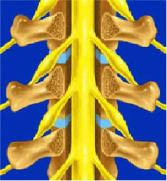
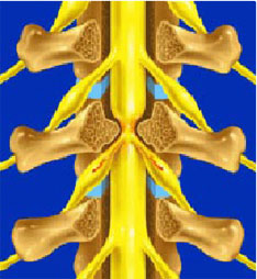
The nerve can move freely in a normal wide spinal canal.
Lumbar spinal stenosis prevents the nerve from moving freely. There is disc bulging and hypertrophy of both the facet joints and the flavum.ligament.
Treatment
If you have severe sciatica, leg numbness or intermittent claudication (=difficulty in long distance walk) and three-month-treatment of spinal injection or other methods fail, Microendoscopic laminoplasty (MEL) is recommended. This surgery can decompress the nerve, so the result is very good and stable.
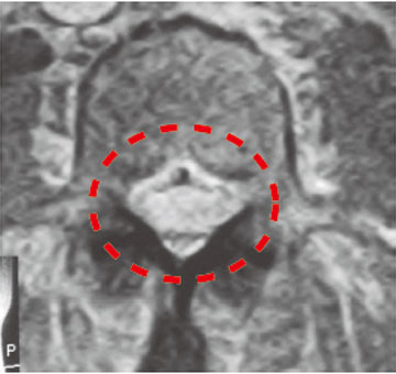
Normal wide nerve on MRI
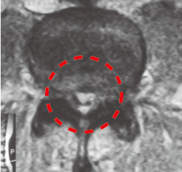
Nerve compressesd in a patient with lumbar spinal stenosis. He has difficulty in long distance walk.
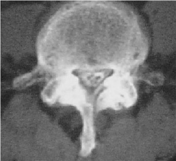
Normal lumbar spinal canal
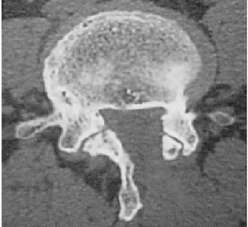
Lumbar spinal stenosis
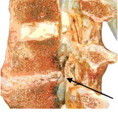
The disc, the flavum ligament, and the bone spur of the facet joint narrow the canal.
MEL(Microendoscopic Laminoplasty)
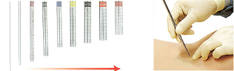
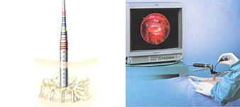
We only use the left two tubes to expand muscle. We used to use all dilators and a 18mm cannula. Now we use only two dilators and the world’s smallest 10mm cannula. The surgery is performed under general anesthesia because the hypertrophied bone which compresses the nerve needs to be resected. It takes about an hour and a patien is required to stay two nights.
Procedure

10 mm cannula is thinner than the index finger
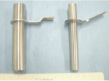
Left: 18mm cannula Right: 10 mm cannula
The nerve is compressed on the bilateral facet joints. This causes leg pain and difficulty in walking.
The canal is decompressed completely. The symptoms go away.